
VETERINARY PHYSIOLOGY
& PHARMACOLOGY
Our department is multidisciplinary and recognized internationally for research in toxicology, reproductive and developmental biology, cardiovascular biology, bone biology, and regenerative medicine.
We do science that matters!
Our goal is to solve major health problems of both humans and animals while providing excellent educational and training programs.
Our integrative scientific approach drives constant innovation and success.
The Department of Veterinary Physiology & Pharmacology (VTPP) at the Texas A&M School of Veterinary Medicine & Biomedical Sciences (VMBS) is committed to a climate where all faculty, students, staff, and administrators feel they are treated respectfully, fully accepted, and thoroughly valued.
In the News
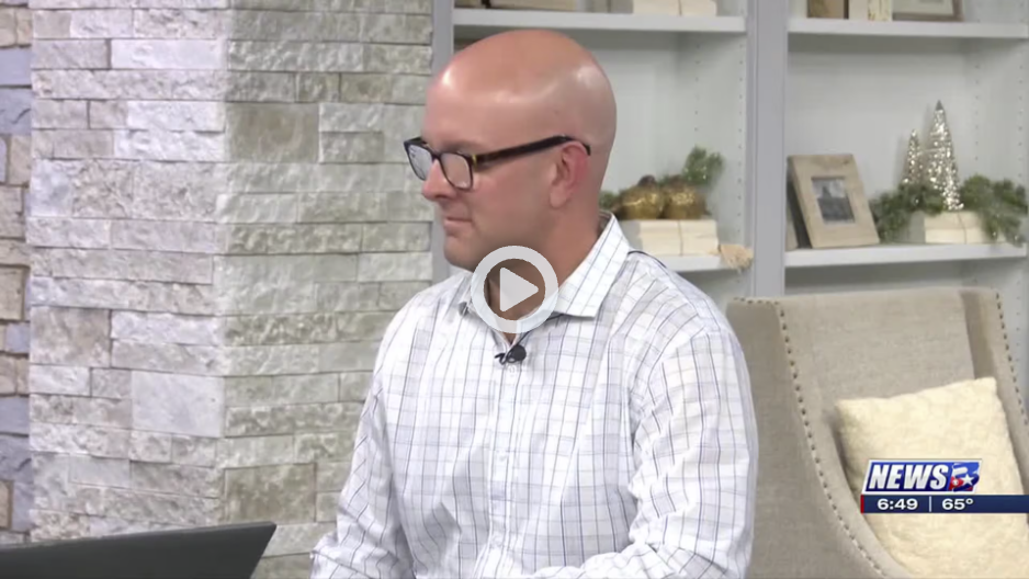
Featuring Dr. Michael Golding
Texas A&M researchers connect father’s alcohol consumption to birth defects (KBTX.com)
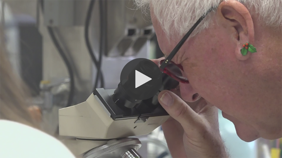
Featuring Drs. Dawson, Porter & Suva
Texas A&M University researchers are studying bone regeneration in individuals with Down Syndrome (KAGSTV.com)
Learn More
Contact Us
Department of Veterinary Physiology & Pharmacology (VTPP)
Texas A&M School of Veterinary Medicine & Biomedical Sciences (VMBS)
4466 TAMU | College Station, TX 77843-4466
Phone: 979.845.7261 | Fax: 979.845.6544